An Ultrasound is a versatile and extremely useful diagnostic medical imaging technique. Performed to view a patient’s internal organs and blood vessels, this procedure is probably best known for being a great way for a pregnant woman and her doctor to check in on the developing baby. However, ultrasounds have many more uses than simply prenatal care.
Doctors use ultrasound images to guide them during minimally invasive procedures. They also use the images produced by an ultrasound to examine the body for problems with muscles, tendons, ligaments, joints, and soft tissue. By using ultrasound, radiologists can assess movement, function, and anatomy in order to analyze a wide range of conditions and determine the extent of damage from injury or illness.
An ultrasound scan also allows doctors to examine the body’s internal organs. The heart, liver, gallbladder, spleen, pancreas, kidneys, and bladder can all be observed, to determine what’s causing pain, swelling, or infection. Ultrasound images are captured in real time, which means they can show the movement of the internal tissues and organs, even letting physicians observe blood flow and heart valve functions.
This diagnostic tool has outstanding versatility and diverse uses. Doctors can use ultrasounds to diagnose damage after a heart attack, locate gallstones and kidney stones, identify fluid cysts, tumors, abscesses, and find impaired blood flow from clots or arteriosclerosis in the legs. These images can also help evaluate superficial structures like the scrotum or thyroid gland.
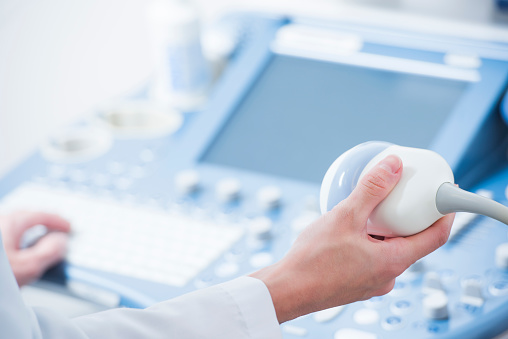
An ultrasound typically takes forty minutes or less. It uses high-frequency sound waves instead of radiation, which is why it’s such a useful obstetrical tool: there’s no risk to the baby from an ultrasound. The typical ultrasound machine consists of a computer, a display screen, and a transducer probe. This probe is the part of the scanner that touches the patient’s body. The care provider will lubricate the area to be scanned, and then place the transducer on top of the place that needs to be scanned.
The transducer produces sound waves, which the body sends back as echoes; yes, an ultrasound works the same way that bats and submarines do. A controlled sound will bounce against any available object, creating an echo that can be used to create an understanding about the shape, size, density, and depth of the item. While bats and submarines use their sonar to navigate, the ultrasound uses controlled sound to create pictures, which are immediately sent to the display screen for the doctor, technician, and sometimes the patient, to view.
For doctors, the benefits of an ultrasound are obvious. Its versatility and ability to create a clear picture of what’s happening to a person internally are extremely valuable and can, in some situations, reduce or even eliminate the need for more invasive procedures. For patients, there are also major benefits: ultrasounds aren’t invasive and they don’t produce unpleasant side effects.
Whatever the reason, if you need to schedule a diagnostic imaging procedure like an ultrasound, Salem Radiology can help. Established in 1974, we are the largest radiology group in the area and offer a depth of specialization among our doctors that you would expect to find only at major university medical centers. To learn more or schedule an appointment, call (503) 399-1262 or contact us through our website.

