Coronary CTA

Did you know that coronary heart disease is the number one cause of death in the United States for both men and women? Nearly a half million people die from coronary heart disease each year in this country, with 250,000 dying before they ever reach the hospital. For about 150,000 of those people, sudden death is the first sign that there’s a problem.

  • Here’s what you need to know about coronary heart disease. Atherosclerosis, a condition in which plaques develop in the walls of the coronary arteries, is usually the cause of coronary heart disease. That’s because if a plaque ruptures, a blood clot can form, restricting the flow of blood to the heart and causing a heart attack.
  • It’s important to know your risk factors. Your risk increases as you age: men over 35 and women over 40 years of age are at a higher risk than their younger counterparts. A family history of heart disease increases your risk, as does a personal history that includes high cholesterol, diabetes, tobacco use, excess weight, or excessive stress. If any of these risks apply to you, it may be time to consider a coronary CTA.  
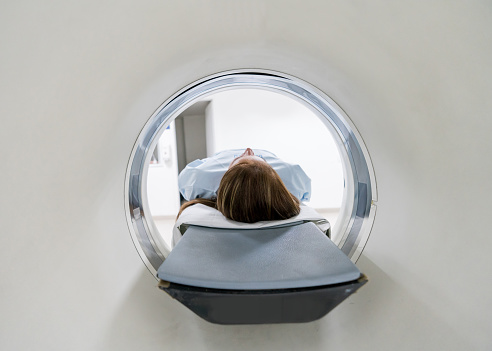
  • What is a coronary CTA? A coronary CT is a non-invasive scan of the heart and chest. It’s quick and painless and allows your doctors to take a look at the calcium build-up in your coronary arteries. This type of scan allows for pictures of the heart to be taken with such precision and quality that heart health can be determined in less than 30 seconds of scan time. It only takes five heartbeats to get images that will allow a Board Certified radiologist, specializing in cardiac disease, to identify your coronary heart health. After thoroughly examining the scan, the radiologist will be able to determine your risk level: 
  • Low risk, with minimal narrowing of the coronary arteries. 
  • Intermediate risk, with moderate narrowing of the arteries that can be controlled by diet, exercise, and medications.  
  • High risk, with severe narrowing of the coronary arteries that requires referral to a cardiologist for immediate surgical intervention.  
  • Talk to your doctor about a coronary CTA. A coronary CTA requires a doctor’s order and is prescribed for patients who have relevant risk factors. It’s a fairly expensive test ($1,195), and its full potential has yet to be determined, but CT scanner technology is advancing rapidly, improving image quality as well as the technique’s accuracy and reliability. It’s an excellent tool for letting doctors know whether a patient is clear from coronary artery disease, or needs further intervention.

If you need a coronary CTA or other diagnostic imaging procedure, Salem Radiology can help. Established in 1974, we are the largest radiology group in the area and offer a depth of specialization among our doctors that you would expect to find only at major university medical centers. To learn more or schedule an appointment, call (503) 399-1262 or ontact us through our website.
Put your Health First in 2019 by Scheduling a Mammogram Screening

Have you been putting off your mammogram? Many women delay having this important screening because it seems like a hassle, or they’re anxious about the procedure, or they just don’t seem to be able to fit it into their schedules. If this sounds like you, it’s time to prioritize your breast health. Make 2019 the year that you put your health first and go ahead and schedule that mammogram.

  • What is a mammogram? A mammogram is a low-dose x-ray examination that can detect breast cancer s and ductal carcinoma in situ (DCIS), an early form of breast cancer. In a mammogram, the breast is compressed so that the radiologist can get a high-quality image of the breast tissue.
  • Why is a mammogram important? Early detection is the best way to beat breast cancer, and a mammogram can find breast cancer far earlier than you or your doctor will be able to feel it. While other types of breast exams, like self-exams, ultrasounds, and MRIs are also useful in detecting cancer, mammography is the only breast cancer screening tool that’s been proven to reduce deaths from DCIS. Mammograms use compression to reduce the dose of radiation necessary, prevent movement, separate the breast tissue, provide uniform thickness, and increase detail in the image, and diminish radiation to the rest of your body. 
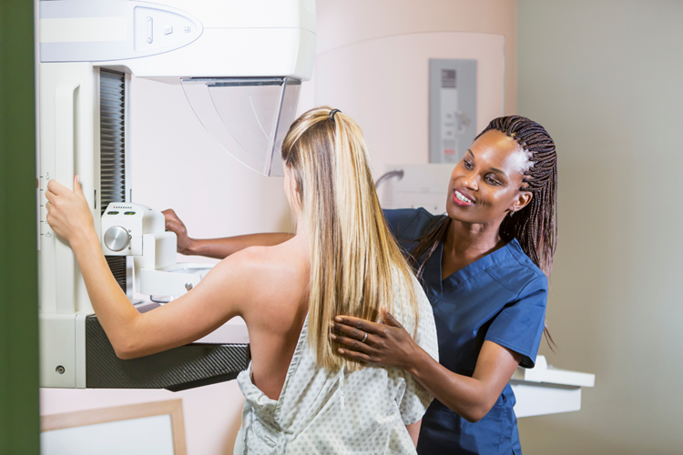
  • What can you expect from your mammogram? A mammogram typically takes about 25 to 30 minutes, including the time you’ll spend filling out paperwork about your medical history. You’ll need to be undressed from the waist up, and a specially qualified radiologic technologist will position each breast on the flat surface of the mammography unit. Your breast will be compressed with a paddle, often made of Plexiglas or another clear plastic, and the technologist will gradually compress your breast and then take images of the breast tissue. Two images are usually needed for each breast, though more may be necessary. While the images are being captured you’ll be asked to hold your breath. The exam may be a bit uncomfortable but typically, that discomfort will quickly subside once the test is over. 
  • What happens next? If there are any questionable areas detected by the mammogram, the radiologist may recommend further imaging tests. You and your referring health care provider will be notified of the results and any further testing or follow-up necessary.  
  • How often should you get a mammogram? For most women, a mammogram is recommended yearly. The age at which you should start getting mammograms is under debate: the American College of Radiology recommends annual screenings beginning at age 40, while the American Cancer Society leaves screenings at the discretion of the woman and her doctor between the ages of 40 and 44, with annual screenings starting at 45. Women with a high risk of breast cancer may need to begin screening much earlier.  Talk to your doctor for advice on what’s best for you.

If you need a mammogram or other diagnostic screening, Salem Radiology can help. Established in 1974, we are the largest radiology group in the area and offer a depth of specialization among our doctors that you would expect to find only at major university medical centers. To learn more or schedule an appointment, call (503) 399-1262 or contact us through our website. 

How Ultrasound Imaging Works

For most people, the term “ultrasound” conjures up the image of excited new parents seeing images of their baby while it’s still inside the mother’s uterus. The truth is, though, ultrasound is an important tool that helps doctors see what’s happening inside a person’s body. While it’s used in pregnancy, it also has a wide range of other uses, including diagnosis, visualization, and even therapy for certain conditions. If your doctor has ordered an ultrasound, here’s what you need to know.

  • Ultrasound imaging works by using sound waves. Through a small transducer placed directly on the skin, the ultrasound transmits high-frequency sound waves into the body. The sounds that bounce back are collected by the transducer and used to create an image of the body’s internal organs in real time.
  • There are different types of ultrasounds. Traditional ultrasounds show images in thin, flat sections of the body, while three-dimensional ultrasounds format sound waves into 3-D images. Doppler ultrasounds, also known as Doppler ultrasonography, allow doctors to see blood flow through arteries and veins. There are three different types of Doppler ultrasounds: color Doppler, which shows the speed and direction of blood flow by using different colors, power Doppler, which is more sensitive than color Doppler but doesn’t show the direction of blood flow, and spectral Doppler, which shows blood flow graphically instead of in a color picture. Spectral Doppler ultrasounds can also convert blood flow information into sound.  
  • Ultrasound technology is non-invasive. It doesn’t require needles or incisions and doesn’t expose patients to radiation the way X-rays and CT scans do. Ultrasounds are painless and have no known harmful effects. They’re also less expensive than many other types of imaging. 
  • Ultrasounds are extremely useful. They can be used to examine the heart and blood vessels, liver, gallbladder, spleen, pancreas, kidneys, bladder, uterus, ovaries, thyroid glands, scrotum, and even babies in utero. In addition to diagnostic use, ultrasound imaging can help guide doctors during procedures like needle biopsies and biopsy of breast cancer.  
  • To prepare for an ultrasound, there’s not much you need to do. For some scans, you may be asked not to eat or drink for twelve hours before your appointment. Sometimes, a doctor will request that the patient drink six to eight glasses of water two hours before the procedure, and avoid urinating to ensure that the bladder will be full. Your doctor will give you specific instructions for your type of ultrasound. The exam usually only takes thirty minutes to an hour.

If you need ultrasound imaging or other diagnostic screening, Salem Radiology can help. Established in 1974, we are the largest radiology group in the area and offer a depth of specialization among our doctors that you would expect to find only at major university medical centers. To learn more or schedule an appointment, call (503) 399-1262 or contact us through our website. 

What is Diagnostic Radiology?

Chances are you’ve heard of radiology which is the science that deals with X-rays and other medical technology that uses high-energy radiation to diagnose and/or treat disease. Not as many people are familiar with the concept of diagnostic radiology though. The word diagnostic simply means the practice or techniques of diagnosis. The term diagnostic radiology refers to a variety of methods that use non-invasive techniques to help identify or monitor diseases.

A variety of tests can be performed using equipment that often involves low doses of radiation. These tests create highly detailed images of areas of the body that are more difficult to diagnose without using diagnostic radiology. This allows the doctors to make a more accurate diagnosis using these detailed images and eliminates the guesswork that is sometimes a result of not being able to see such detail.

The science of diagnostic radiology can be used to identify many different medical conditions including heart conditions, blood clots, broken bones, gastrointestinal conditions, and more. Medical professionals can also utilize diagnostic radiology to monitor how your body responds to a treatment you are currently undergoing. The technology is also used to screen for certain diseases like breast or colon cancer.

There are different types of machines used in diagnostic radiology, but the most common include:

  • X-ray machine – Utilizes X-rays which are a type of electromagnetic radiation and produces images of the interior of your body without any incisions being made.
  • CT scanner – This machine utilizes X-ray equipment and creates a sequence of cross-sectional images of your body. This procedure is more common when the doctor requires highly detailed images.
  • MRI machine – This machine utilizes a magnetic field and produces images of the inside of your body. It’s most often used for bones or other areas of the body that CT scanners are not as effective on.

To learn more about diagnostic radiology and how it can help you, contact Salem Radiology at (503) 399-1262.

Do I Need An MRI?

MRIs are a popular way for a doctor to get high-quality images of your spine in order to come up with an effective treatment for back pain, but how do you know if you need an MRI? The term MRI is an acronym for Magnetic Resonance Imaging. The technology was introduced in the 1980s and has helped millions of people who live with lower back pain.

An MRI is most commonly used for pre-surgical planning to determine if a patient would benefit from a decompression or a lumbar spinal fusion procedure. MRI scans are very thorough and provide detailed information including the health of the discs in your back and the possible presence of a lumbar disc herniation or any spinal tumors.

MRIs can also rule out an infection or tumor, differentiate scar tissue from recurring disc herniation for patients who have had back surgery, and rule out the risk of injecting a steroid into an infection or tumor before performing an epidural injection. While an MRI is often beneficial for patients who have lower back pain or leg pain, it’s not always the right option.

There are many factors that need to be considered before someone undergoes an MRI scan. While many patients do benefit from getting an MRI, it’s sometimes best to wait a certain period of time because sometimes back pain will get better, and the procedure can be expensive. MRIs can sometimes detect problems that are not currently causing you any pain which can confuse the matter.

The reality is that MRIs can be very helpful and are used in many patients with back, neck, or leg pain. Chances are you aren’t really going to know if you need an MRI unless you are a medical professional. Your doctor is the one that will ultimately make the decision of whether or not you need an MRI based on their professional opinion. If you think you might need an MRI, the first thing to do is to schedule an appointment with Salem Radiology by calling (503) 399-1262 today.