Common Mammogram Myths

If you’ve been told you need a mammogram, you may have questions. There’s a lot of misinformation circulating, and some of it may lead you to believe that you shouldn’t get a mammogram because it’s not important or even dangerous. Here, we look at some common mammogram myths, so that we can debunk them with the truth.

  • Myth #1: Mammograms aren’t important if you have no family history of breast cancer. Whether or not you or anyone in your family has ever had symptoms of breast cancer, if you’re over 40 years old you should be getting an annual mammogram. Mammograms are used for early detection, which means they find cancer before you can even feel a lump. When you catch breast cancer in the early stages, your chances of survival are significantly higher than if you wait. How significant? Early detection leads to a survival rate of 99 percent, as opposed to late-stage discovery of breast cancer, which gives you only a 27 percent chance of survival. Consider this fact: 75 percent of women with breast cancer have no family history of the disease.
  • Myth #2: The radiation used in mammograms is unsafe. It’s true that mammograms use radiation, but it’s a very small amount. Mammography is a highly regulated screening tool, under the supervision of the Food and Drug Administration and other regulating agencies. As long as you go to a facility certified by these agencies, there’s no cause to question the safety of the procedure. In fact, the dose of radiation that you receive from a mammogram is about the same as radiation you’d get from just living everyday life for two months. 
  • Myth #3: A traditional mammogram is just as good as a 3-D mammogram. The 3-D mammogram is much more advanced than the traditional 2-D mammogram, displaying more images of the breast that can be viewed by the physician radiologist from different angles and depths. This provides greater clarity, which allows doctors to more accurately distinguish between normal tissue and cancer. The most modern diagnostic tool available for early detection of breast cancer, the data provided by 3-D mammography is 40 percent more effective in detecting early cancer, with a 40 percent decrease in false alarms. 
  • Myth #4: A screening mammogram will find any type of cancer present in breast tissue. There are limitations to mammograms, and when breasts are very dense, cancer is often hidden by the tissue. Sometimes, normal breast tissue doesn’t just hide cancer, it mimics it. That’s why doctors additionally use other tools such as breast ultrasound or a breast MRI, for women with dense breast tissue. 
  • Myth #5: If your mammogram is normal, you can skip the next year’s mammogram. Mammograms are not preventative, so a clear mammogram does not guarantee that future mammograms will be normal. When you have a mammogram every year, you increase the chances that any cancer will be detected when it’s small and easily treatable. 
  • Myth #6: A referral from your doctor is necessary for a mammogram. It is preferred that you receive an order from your provider in coordination with a physical breast exam; however,  the federal government allows women the option to self refer for a screening mammogram.  A referral from your doctor is not absolutely necessary but is recommended.  It’s smart to be proactive with your breast care and schedule a mammogram every year. 

If it’s time for you to schedule a mammogram, Salem Radiology can help. Established in 1974, we are the largest radiology group in the area and offer a depth of specialization among our doctors that you would expect to find only at major university medical centers. To learn more or schedule an appointment, call (503) 399-1262 or contact us through our website.
The Stages of Vein Disease

Could you be suffering from vein disease? Millions of Americans ignore the warning signs of vein disease, only seeking treatment when complications like venous leg ulcers arise. It’s important, however, to seek vein disease treatment as soon as possible, to protect your vascular health and prevent the disease from worsening over time.

  • Are you at risk for vein disease? There are several risk factors for vein disease, and in fact, recent research from Stanford Medical School found 30 genes that may be associated with varicose veins. The most common risk factors for vein disease include:
  • Being age 50 or older
  • Pregnancy
  • Overweight
  • Prolonged sitting or standing
  • Smoking
  • Family history of vein disease
  • How can you tell if you’re in the early stages of vein disease? Assess yourself. Do you notice spider veins forming, and do your legs feel heavy when you’re standing up, but better when you’re resting? If so, see a vascular specialist. Vein disease starts with subtle changes like spider veins, which look like scattered or tangled clusters of wispy red or purple lines. They’re flat, and when you rub your hand over them, you won’t feel bulging or swelling. Another early sign of vein disease is varicose veins, which are bulging, ropy, and often quite painful. Varicose veins contribute to leg pain, itching, tingling, swelling, restlessness, and heaviness in the legs, and these symptoms are often the reason people seek medical intervention. 
  • What happens in the later stages of vein disease? When people with vein disease delay seeking medical attention, the disease can progress to a point that significantly impairs quality of life. In the later stages, it may be more difficult to relieve symptoms by resting your legs or using over-the-counter medications. Varicose veins can lead to skin discoloration and leg swelling because then the veins in the legs are not functioning properly, the body can’t absorb fluid. It’s also easy, at this stage of vein disease, to develop open sores that may recur or take a long time to heal. Venous leg ulcers often come next, because of inadequate blood flow bringing nutrients to the skin. These ulcers can become chronic, painful, and debilitating, and may also introduce bacteria into your legs, causing pain and increasing the risk of infection. Left untreated, vein disease can cause significant disease to the lymphatic system, resulting in lipid accumulation and further damage to the veins. 
  • What can be done about vein disease? Vein disease treatment varies depending on the stage of the disease. Compression stockings may help keep blood flowing properly, and topical or oral medications can fight infection or inflammation. Your doctor may decide on sclerotherapy, which involves injections into affected veins to reroute blood flow, or microphlebectomy, in which the problematic vein is removed through a small nick in the skin. Endovenous thermal ablation is another option, using high-frequency radio waves or heat from a laser to collapse the problem vein inside of itself, which is then reabsorbed by the body. The best time to seek treatment is in the early stages of vein disease, but these treatments can restore your quality of life even in the later stages. 

If you’re concerned about your legs or have noticed the first symptoms of vein disease, Salem Interventional Radiology can help. Established in 1974, we are the largest radiology group in the area and offer a depth of specialization among our doctors that you would expect to find only at major university medical centers. To learn more or schedule an appointment, call (503) 399-1262 or contact us through our website. 

What to Expect at Your First Mammogram

Have you been putting off getting your first mammogram? For most of us, the thought of a mammogram is a little intimidating. You’ve probably heard a variety of jokes or stories related to the breast compression that occurs with a mammogram. The truth? Mammograms save lives, and that’s why they’re recommended annually for women age 40 and over. It’s important to try not to be anxious, and knowing what to expect from your first mammogram appointment may help to settle your nerves, reduce your stress, and give you more confidence.

First, let’s talk about the technical aspects of a mammogram. Mammograms use low-dose x-rays to examine breast tissue and help doctors detect abnormalities. Research has shown that mammograms find lumps that may be too small to be felt, which means cancer is caught in the very early stages when it’s most treatable. This is important because when cancer is treated early, patients tend to have much better outcomes.  

During the mammogram, there’s a pretty standard procedure that you can expect to experience.  

  • First, you’ll undress. You’ll be given a gown, and asked to undress from the waist up. Don’t wear anything scented on your body, like deodorant, lotions, oils, or perfumes, because this can interfere with the machine. You don’t want to have to come back because your lotion affected your result!
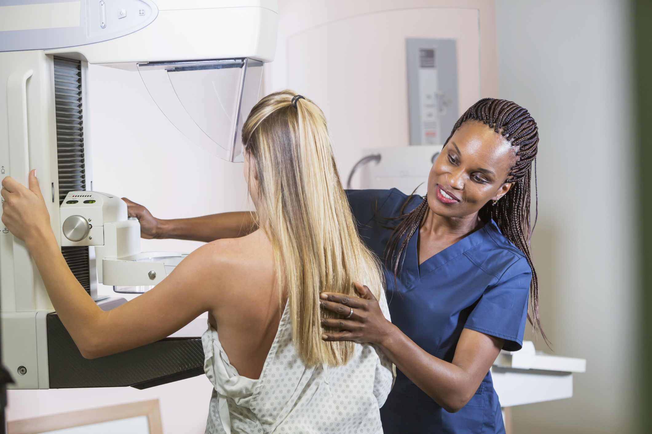
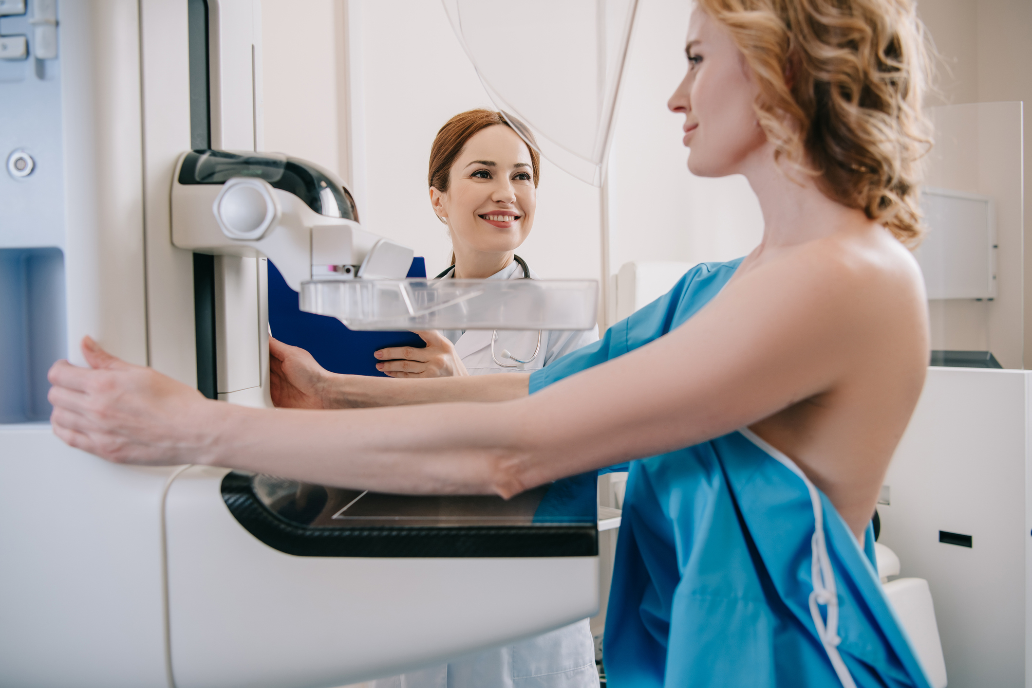
  • Once you’re in your gown, you’ll head into the exam room. In this room, it will just be you and a trained female technologist. The technologist will help you position your breasts one at a time between two plastic imaging plates, and then will take multiple images from different positions.
  • You’ll feel pressure on your breasts. The plastic imaging plates apply pressure, which spreads out the tissue so that clearer images can be captured. Because the breast tissue is spread out, less radiation is required for the imaging. This process, however, can be uncomfortable, and it’s what patients typically dread about having a mammogram. The compression; however, should never be painful.
  • This too shall pass. It’s human nature to tense up when we’re anxious about something, but it’s really important to stay relaxed so that your muscles don’t work against the machine. The whole process, including both breasts, only takes about ten minutes, so it’s not too difficult to just breathe deeply and get through it. Most patients are pleasantly surprised, and many say that the process wasn’t as bad as expected.
  • The type of mammogram determines the speed of the results. If a diagnostic mammogram was ordered because of a change in symptoms, the radiologist will review your scans while you’re there. That way if additional imaging is required, it can be requested right away. If it’s a screening mammogram, the results will usually get to your doctor in about 48 hours.

If it’s time for you to schedule a mammogram, Salem Radiology can help. Established in 1974, we are the largest radiology group in the area and offer a depth of specialization among our doctors that you would expect to find only at major university medical centers. To learn more or schedule an appointment, call (503) 399-1262 or contact us through our website.  
What’s the difference between an MRI and a CT Scan?

If you’ve been scheduled for diagnostic imaging, you probably have questions. The first time you have any medical procedure, it can be a little bit daunting, but don’t worry! MRI and CT scans are non-invasive and non-surgical, and there’s very little discomfort involved. What’s the difference between these two types of tests? We’ll break it down for you.

  • MRI stands for Magnetic Resonance Imaging. An MRI machine uses superconducting magnet and radiofrequency waves to provide detailed information about the inside of a person’s body. This kind of test is generally performed when doctors want to know more about the brain, skeletal system, reproductive system, and other organ systems. There’s no preparation necessary for an MRI, and while the machine is noisy and can make some patients feel claustrophobic, the test itself is not invasive or uncomfortable. MRIs take about 20 minutes for each system the doctor wants to examine.
  • A CT scan is a Computerized Tomography scan. Using a sophisticated x-ray machine, a CT takes multiple detailed pictures of the inside of the body and transfers those images to a computer. The images can then be viewed from different directions and angles or rotated in a three-dimensional model. The procedure is painless, but in some cases, a dye or contrast material is used to make blood vessels or organs easier to see. Sometimes this dye is injected, and sometimes it’s swallowed. CT scans are much quicker than MRIs, requiring only about 20 seconds of actual scan time for each part of the body being scanned. There is some concern about the long-term radiation exposure risks from CT scans, but at Salem Radiology, we offer a “low dose” option, which provides high-quality images with the lowest possible radiation.

Why would a doctor prefer one of these tests over another? One consideration is that in addition to taking a much longer time, an MRI is much more expensive than a CT scan. On the other hand, MRIs provide much more detail about soft tissue than CT scans, so if there’s a question upon review of the CT, a doctor might decide to order a follow-up MRI. Talk to your doctor about your options, and make sure you’re comfortable with the tests being ordered for your care. 

Whether you need an MRI, a CT scan, or some other form of diagnostic imaging, Salem Radiology can help. Established in 1974, we are the largest radiology group in the area and offer a depth of specialization among our doctors that you would expect to find only at major university medical centers. To learn more or schedule an appointment, call (503) 399-1262 or contact us through our website. 

The History and Advancement of Mammograms and Mammography

Mammograms are the most well-known form of breast imaging, so most people have at least a passing familiarity with what they are and how they work. How much do you know about mammography? Would it surprise you to know that radiologists have been performing mammography exams since the 1960s? The history of mammography reflects an ever-evolving scientific pursuit by medical professionals dedicated to improving women’s healthcare.

In the early days, mammography was performed using x-ray machines. In fact, the idea of using x-rays to examine breast tissue dates back to 1913, when a German surgeon began studying breast tissue removed in mastectomies to see if it was possible to identify cancer through those images. Finding microcalcifications that correlated to known cancer pathology, he wrote about the usefulness of x-rays in identifying cancerous tumors. As early as 1949, the compression technique was being used in breast imaging, and that technique is still being used today. It was in 2000 that the FDA approved the first digital mammography unit, and 11 years late the first 3D breast imaging technology was approved.  

Today, 3D mammography remains the standard of care. More accurate than traditional 2D mammograms, 3D mammograms allow for better breast detection, which means a lower likelihood that additional screenings will be necessary. 3D mammography can detect breast cancer 15 months earlier and identify 41 percent more invasive cancers than conventional mammography. 

Other methods of breast cancer screening use magnetic resonance (MRI) technology. Breast MRIs have been recommended by the American Cancer Society for women who are at high risk of developing breast cancer, either because they are known carriers of the BRCA1 and BRCA2 genes or are first-degree relatives of someone with either of those gene abnormalities. Breast ultrasounds are used to further characterize suspicious masses previously detected with mammography, and ultrasound-guided breast biopsies are a minimally invasive, highly accurate tool for determining whether an abnormality is cancerous or benign.

At our practice, a radiologist certified as a specialist in the interpretation of breast images reads diagnostic mammograms immediately after the exam, to determine if additional imaging is needed before the patient even leaves the facility. We also utilize a sophisticated computer analysis system known as computer-aided detection, or CAD, to provide a “computerized second opinion” for every mammogram performed at Salem Radiology Consultants. After reviewing the films thoroughly, the radiologist consults the CAD system for input into the final report, a practice shown to increase the early detection of breast cancer by as much as 19 percent.  

If it’s time to schedule a mammogram, Salem Radiology can help. Established in 1974, we are the largest radiology group in the area and offer a depth of specialization among our doctors that you would expect to find only at major university medical centers. To learn more or schedule an appointment, call (503) 399-1262 or contact us through our website.