Treatment Options for Varicose Veins

The mildest form of varicose veins, known as spider veins, may not require a doctor’s care. However, as vein disease progresses, you may need treatment to relieve the resulting discomfort, redness, swelling, and ulcers. Consider how vein disease progresses and the best options for treating varicose veins at home or with a doctor’s help.

The Progression of Vein Disease

Stage 1: The first stage of vein disease is called spider veins. When these first develop, you may notice the appearance of dilated skin capillaries just under the skin. These create a spider-like appearance, but they usually cause no further symptoms.

Stage 2: If spider veins progress to varicose veins, they become rope-like and bulge from the surface of your skin. This is the stage of vein disease when you may begin to experience achy, tender, and sore legs, which may interfere with your day-to-day life.

Stage 3: Left unaddressed, varicose veins may evolve into other health problems, such as leg edema. This condition is characterized by chronic swelling that worsens as the day goes by and after standing for long periods. At this stage, some people also experience restless leg syndrome, itchy skin, and leg cramps.

Stage 4: Over time, vein disease can lead to skin changes, including discoloration and thinning of the epidermis. It may begin to appear as though you have brown stains on your skin, which occurs as blood leaks from your blood vessels into the surrounding soft tissue.

Stage 5: In the final stage of vein disease, leg ulcers develop. These may be extremely painful and itchy, requiring constant care and dressing to avoid infection.

At-Home Treatments for Varicose Veins

In the early stages of vein disease, you may not be too concerned if you’re not in any pain. However, even if there’s no discomfort, you shouldn’t ignore superficial spider veins and newly developing varicose veins. Prompt care may improve their appearance, but what’s more, at-home treatment can prevent the progression of vein disease. Here’s what you can do without additional assistance from a doctor.

Wear Compression Stockings

You can find over-the-counter stockings at a pharmacy or medical supply store. Expect mild to moderate compression from these products. Higher pressure stockings require a prescription. The purpose of compression stockings is to help your muscles push blood up your leg. They provide the most compression at the ankles and gradually decrease in pressure the higher up your leg they go.

Put on your compression stockings in the morning before getting out of bed. While lying on your back, raise your legs into the air and pull the stockings on evenly. Wear them all day, if possible. Also, take breaks from your daily activities to elevate your legs for 10 to 15 minutes several times a day.

Take OTC Anti-Inflammatory Drugs

Aspirin and ibuprofen are non-steroidal anti-inflammatory drugs (NSAIDs) designed to reduce inflammation and treat mild to moderate pain. You can take these over-the-counter medications with or without wearing compression stockings.

Medical Procedures for Varicose Veins

At-home care is often enough to prevent spider veins from getting any worse. Still, you should contact a doctor immediately if you notice the skin around a varicose vein becoming discolored or ulcerated. You may also need medical treatment if you experience vein-related leg pain, even if there are no obvious outward symptoms.

The goal is to treat your vein condition with the least invasive procedure possible. Consider your options.

Interventional Radiology

This minimally invasive treatment is an alternative to open surgery. It involves placing a catheter inside the body with the aid of image-guided technology, such as CT scans, X-rays, or MRIs. Heat is then applied with radiofrequency waves or lasers to destroy and ultimately close the varicose vein.

Laser Treatment

This medical procedure is usually performed on small varicose veins. It involves directing light energy from a laser at a bulging vein, causing it to gradually fade and disappear. Multiple treatments may be needed to see the final results.

Sclerotherapy

Small to medium-sized varicose veins may be treated with sclerotherapy. This chemical treatment involves injecting material into the affected vein, causing it to harden and shrink so it can no longer transport blood.

Surgery

As a last resort, surgical removal—also known as ligation and stripping—may be used to treat large varicose veins that have advanced past the early stages of vein disease. Be aware that surgery may leave tiny scars behind.

Schedule a Free Vein Screening in Salem, OR

Salem Radiology Consultants is proud to be the largest radiology group in the Salem, Oregon area. Our depth of specialization ranks among the services you would expect to find at major university medical centers. If you’re concerned about bulging, uncomfortable veins in your legs, we can help. Contact us at (503) 399-1262 to schedule a free vein screening at our clinic today.

The Benefits of 3D Mammography

Early detection is the key to winning the fight against breast cancer. That’s why the American Cancer Society recommends annual breast cancer screenings starting as early as age 40. Different tests are available to help find cancer before you exhibit any outward symptoms. Mammograms are the standard screening method.

There are two types of mammograms. First is the conventional version, which takes a two-dimensional image of each breast to look for any abnormalities. The second more advanced breast imaging technology is called 3D mammography. This uses several x-ray images taken from different angles to create a three-dimensional picture.

3D mammography is FDA-approved, but is relatively new, and isn’t offered at all hospitals and mammogram facilities yet. Salem Radiology Consultants is pleased to offer 3D mammography to our patients in the Salem and the surrounding communities .

How Does3D Mammography Work?

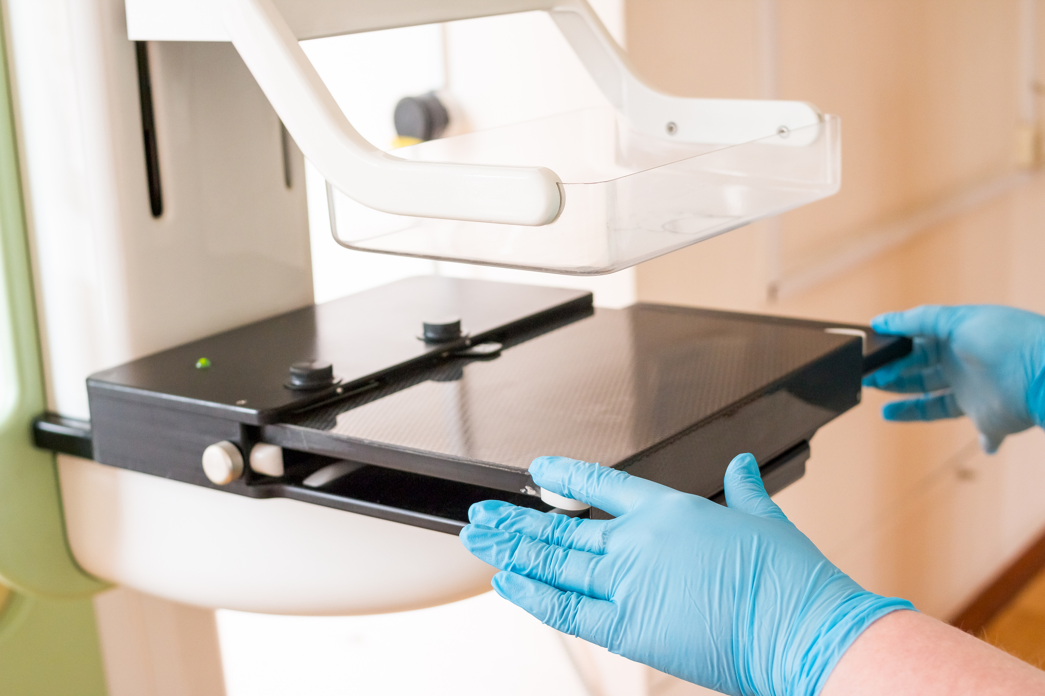
When you visit our clinic for a 3D mammogram, a female technologist helps to position you in front of the machine, which gently compresses your breast to hold it stable during the procedure. Then, an X-ray tube moves in an arc across the breast, capturing 11 images in just seven seconds. The information is sent to a specially designed computer where a physician radiologist carefully examines the highly focused, 3D images of your breast tissue.

Why Choose 3D Mammography?

Consider the benefits of this superior breast screening technology as outlined in Journal of the American Medical Association (JAMA).

Reduced Callbacks

If the doctor detects abnormalities in your mammogram, you may be called back to perform additional imaging. 3D mammography reduces callbacks by 40% eliminating the stress, time, and additional costs associated with follow-up imaging and potential biopsies.

Enhanced Cancer Detection

Screening with 3D mammography leads to a 41% increase in invasive cancer detection compared to conventional mammograms.

Earlier cancer detection

A 2D screening is akin to standing at the edge of a forest and looking for a bird somewhere inside. A 3D screening, on the other hand, is like walking into the forest and looking around 10 steps at a time. The more in-depth nature of 3D imaging allows doctors to detect breast cancer an average of 15 months earlier, giving patients a critical head start in the battle against cancer.

Computerized Second Opinion

The computers used in 3D mammography employ a sophisticated analysis system called computer-aided detection (CAD). After examining the images closely, radiologists consult the CAD system before issuing their final report. This combination of human interpretation and computer analysis increases early breast cancer detection by as much as 19%.

Improved Images in Dense Breast Tissue

Breasts consist of milk glands, milk ducts, and supportive tissue. Breasts are considered “dense” if they contain more supportive tissue than fatty glands or ducts. Dense tissue and cancer both appear as white spots on a standard mammogram, which makes cancer more difficult to detect in dense breasts. However, with 3D mammography, doctors can see beyond areas of density from a different angle, making it easier to detect the presence of cancer and reducing the number of false positives.

More Comfortable Screening

The breast compression required during a conventional mammogram is uncomfortable for many women, causing some women to delay scheduling their exam. Compression also causes some tissue to overlap, giving breast cancer a place to hide. 3D mammography machines only apply enough pressure to keep the breast in a stable position. This makes the procedure much more comfortable without detracting from its effectiveness.

According to one of the senior authors of the study published in JAMA, “These findings reaffirm that 3D mammography is a better mammogram for breast cancer screening. These results are an important step leading toward updated policies so that all women can receive 3D mammography for screening.”

Limitations of 3D Mammography

While this technology provides the safest, and most effective way to screen for breast cancer, it does have a few technological limitations that patients should know about.

Low-Level Radiation Exposure

3D imaging using multiple x-rays to create a three-dimensional image, which exposes patients to low levels of radiation. 2D mammograms use x-rays as well, but because 3D screenings are often combined with standard mammograms, the overall radiation exposure may be slightly higher than using 2D technology alone. However, Salem Radiology’s 3D machines create both two- and three-dimensional images at once, lowering the amount of overall radiation.

Unable to Detect All Cancers

While 3D mammography is better at finding cancerous growths earlier than 2D screenings, small or difficult-to-see tumors may not be detected by the technology. That’s why, no matter what type of imaging you choose, you should continue to schedule breast cancer screenings every year and visit your doctor for a physical breast exam.

Schedule 3D Mammography in Salem, OR

Salem Radiology Consultants is committed to offering the best medical screenings and diagnostic services to our patients. As such, we are proud to be among the early adopters of 3D mammogram technology.

In addition to 3D mammography, we also offer additional imaging techniques, including breast MRIs, breast ultrasounds, and ultrasound-guided breast biopsies. Whether you need preventative screenings or diagnostic services, we can meet your needs at our imaging facility.

If you still have questions, or you’re ready to schedule a 3D mammogram, please contact Salem Radiology Consultants at (503) 399-1262.

Differences Between an MRI and a CT Scan

When you have a medical issue that requires diagnostic imaging, your doctor may recommend an MRI or CT scan. Both are non-invasive, low-risk ways of finding out what’s going on inside your body. One test might make more sense than the other, depending on your situation.

What is an MRI?

MRI stands for magnetic resonance imaging. It uses a magnetic field and radio waves to capture detailed images of the structures inside your body. MRIs are typically used to diagnose issues with the brain, heart, blood vessels, breasts, spine, and joints.

An MRI machine works by bouncing a constant magnetic field and pulsing radio frequencies off of the fat tissue and water molecules in your body. Radio waves transmit to a receiver, which translates them into images of the body that your doctor can use to diagnose health problems.

What is a CT Scan?

CT stands for computed tomography. You may have also heard of a CAT scan, which stands for computer axial tomography. This technology is one and the same; CT scan is simply a more modern term for CAT scan.

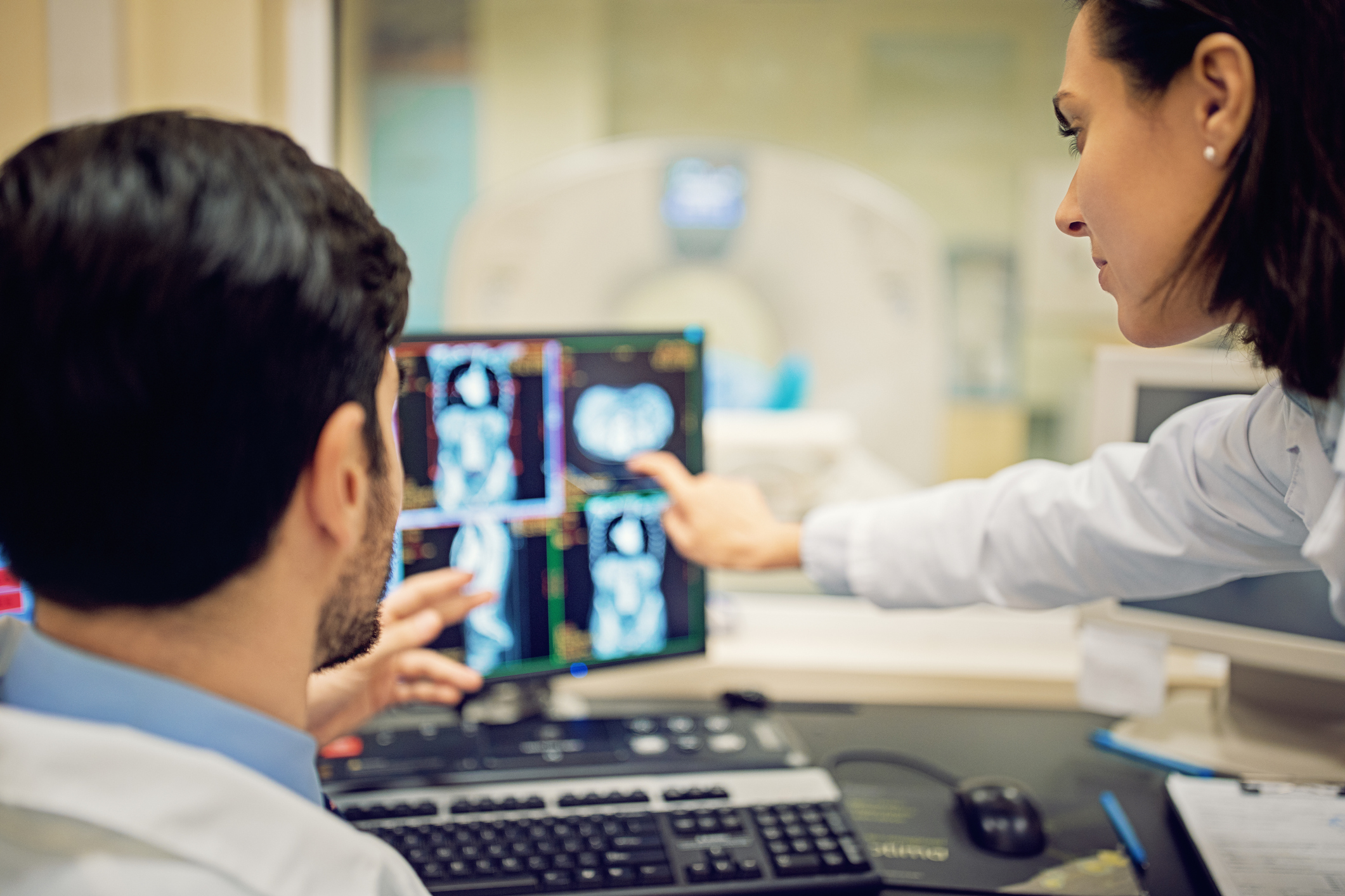
CT scans are a type of x-ray that captures multiple detailed pictures of the inside of your body, one layer at a time. A sophisticated x-ray machine transfers those images to a computer, which arranges each cross-sectional picture into a rotatable 3D model. CT scans are effective for diagnosing bone fractures, internal bleeding, tumors, and cancer.

MRI vs. CT Scan

Technology & Costs

To the untrained eye, MRI machines and CT scanners look very similar. They both have donut-shaped holes with a scan table sticking out of them. However, the technology used to achieve the scans is quite different.

MRIs are considered the superior option because magnets and radio waves provide more detailed imaging than x-rays. However, MRIs are more costly, and the scan takes about 20 minutes for each body area being imaged.

CT scans, which are the less expensive option, are more widely used than MRIs. They are also fast, taking less than 20 seconds to scan you from head to toe once you’re situated on the scan table. Be aware that some procedures require two passes across the body, which doubles the scan time.

Risks

While both imaging methods are considered safe, there are some risks involved.

MRIs have the following risks:

  • Possible feelings of claustrophobia
  • Chance of increased body temperature during long procedures
  • Potential adverse reaction to any metal in the body (consult a doctor before receiving an MRI if you have artificial joints, an IUD, eye implants, or a pacemaker)

CT scans have the following risks:

  • Harm to unborn babies
  • Exposure to a low dose of radiation, just like ordinary x-rays
  • Potential reaction to the dyes sometimes used to enhance the scan

Preparation & Procedure

What to expect from an MRI:

No preparation is required for an MRI. For your safety, simply tell your doctor if you are pregnant or have implants of any kind.

You must lie still while receiving an MRI. Since the procedure is much longer than a CT scan, this may be challenging for some patients.

Rest assured that an MRI is painless. You don’t feel the magnetic field or radio waves passing through your body, and there are no moving parts around you. However, MRI machines are loud, with repetitive tapping, thumping, and other noises. You may be offered earplugs or headphones to make the procedure more pleasant.

What to expect from a CT scan:

If you receive a scan without contrast materials, minimal preparation is necessary. Plan to wear comfortable, loose-fitting clothes that contain no metal zippers or snaps. You have the choice to change into a medical gown if you wish. You should also remove any body piercings, jewelry, glasses, and metal hair accessories.

In some cases, a dye or other contrast material is needed to make your blood vessels and organs easier to see in the x-ray images. If your CT scan requires this, be prepared to have an intravenous (IV) line gently inserted into your arm before the scan begins.

While the scan is in progress, you will hear clicking and whirring while the machine rotates around you, but there is no pain, and nothing comes in contact with your skin. Note that patients must weigh 500 pounds or less to receive a CT scan due to the scan table load limit.

Make the Most of Your MRI or CT Scan

Salem Radiology Consultants offers the most advanced MRI technology known as 3T. A “T,” or Tesla, is a measurement of the magnetic field strength. Before the introduction of the 3T machine, the standard was 1.5T.

Greater magnetic strength means our MRIs provide improved image quality with high-definition clarity. Scans also take less time with stronger magnets. In addition, our 3T MRI machine features a larger opening, making the scanning experience more pleasant, especially for patients with claustrophobia. There is no extra cost for receiving a higher-quality 3T scan, so it makes sense to take advantage of the best technology available.

Our CT scanner is equipped with advanced ASIR technology whichperforms scans with the lowest possible dose of radiation for drastically reduced radiation exposure and greater safety to our patients. Rest assured that the results are superior, so there’s no reason not to choose the low-dose option.

If you have any questions about which type of diagnostic imaging is right for you, or you’re ready to schedule your MRI or CT scan, please contact Salem Radiology Consultants at (503) 399-1262.

What is a Bone Density Scan?

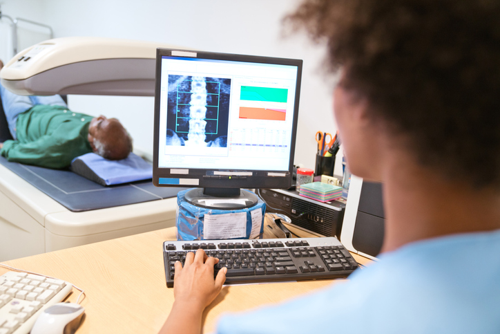
If your doctor suspects that you might have osteoporosis, you’ll probably be sent for a bone density scan also known as a DEXA scan. Osteoporosis occurs when your bones become thin and weak because they’re lacking in calcium and other important minerals. Osteoporosis, when treated by your physician, can significantly lower your risk of serious fractures.  To help evaluate whether or not you have osteoporosis, your physician may recommend a bone density test (DEXA scan) to measure the strength of your bones and your risk of fracture.

  • Several different factors might lead a doctor to recommend a bone density scan. Doctors use these scans to identify a decrease in bone density before any bones actually break, and determine whether or not you’re at risk for fractures. Osteoporosis is most common in post-menopausal women, but people of any gender and age can be affected by it. Therefore, regardless of your age or sex, your doctor may recommend a bone density scan if you have:
    • Lost at least 1.6 inches in height.
    • Broken a bone with only mild trauma.
    • Taken certain drugs, like steroids.
    • Received an organ or bone marrow transplant.
    • Had a drop in hormone levels, whether it’s due to the natural aging process or because of cancer treatments.
  • The scan is easy, quick and painless, and doesn’t require much preparation. You’ll need to tell the doctor if you’ve had any tests with contrast, because that can interfere with your bone density test, and you’ll also need to avoid calcium supplements for at least 24 hours before your bone density test. Wear loose, comfortable clothing, avoiding any clothes with belts, zippers, or buttons, and removing all metal objects from your pockets. The test only takes 20-30 minutes, during which time you’ll lie on a padded platform while a mechanical arm passes over your body. A bone density test doesn’t expose you to very much radiation; it’s less than the amount emitted during a chest x-ray.
  • Understanding the results of the scan is a little bit tricky. The higher your bone mineral content, the denser your bones are, and the less likely they are to break. The results are reported in two numbers: the T-score and the Z-score.
    • The T-score compares your bone density with the bone density expected in a healthy young adult of your same gender. It’s expressed in numbers that represent how far above or below average your bone density is. A score of -1 or above is normal, while a score between -1 and -2.5 is indicative of osteopenia. Osteopenia is a condition that involves low bone density, and it can lead to osteoporosis if left untreated. If your result is below -2.5 you likely already have osteoporosis.
    • The Z-score indicates your deviation above or below what’s expected for someone of your age, sex, weight, and racial or ethnic origin. If this score is much higher or lower than the average expected number, something other than aging may be responsible for your abnormal bone loss. In this case, the doctor will attempt to identify the underlying problem, so that the condition can be treated. The right treatment can slow or even stop bone loss.

If you need a bone density scan, contact Salem Radiology. Established in 1974, we are the largest radiology group in the area and offer a depth of specialization among our doctors that you would expect to find only at major university medical centers. To learn more or schedule an appointment, call (503) 399-1262 or contact us through our website

How to Prepare for an Ultrasound

Has your doctor recommended an ultrasound? A diagnostic ultrasound uses high-frequency sound waves to create images of organs and other structures inside your body. Ultrasound imaging, also known as sonography or diagnostic medical sonography, is a valuable tool for diagnosing and treating a wide range of diseases and conditions.

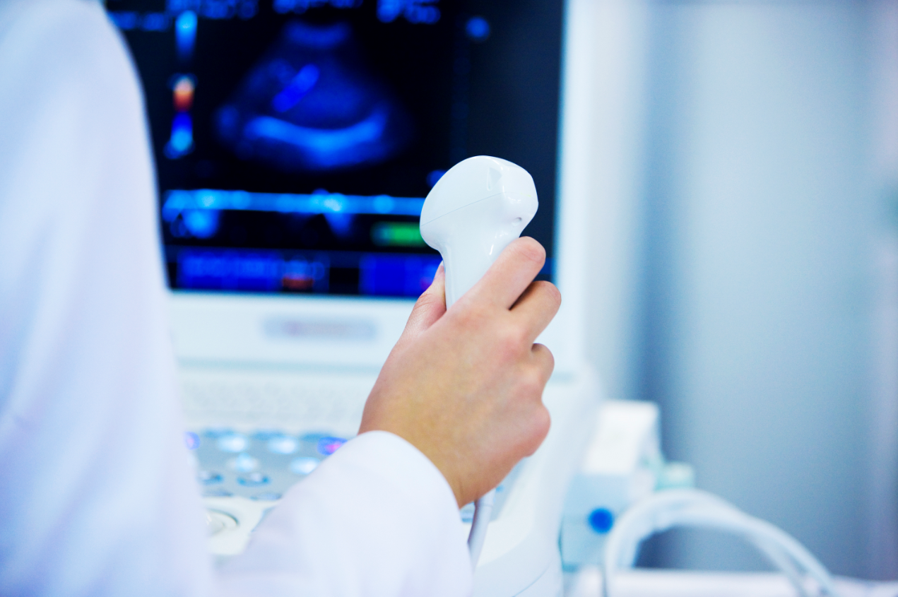
During an ultrasound, a sonographer will use a handheld wand or probe to send sound waves into your body. Typically, the ultrasound device is outside of your body during the procedure, but some ultrasounds involve placing it inside your body. The sound waves are reflected back to the machine in a way that creates two and three-dimensional images, allowing the radiologist to examine images from the targeted area. Ultrasound scans can be used in obstetrics and gynecology, cardiology, urology, in the diagnosis of cancer, and to evaluate orthopedic conditions. Ultrasound doesn’t use radiation, and it’s less expensive than some other forms of imaging.

There’s not much preparation for an ultrasound. What’s required depends on the type of ultrasound you’re undergoing.

  • For the abdomen: If your liver, kidneys, gall bladder, biliary ducts, pancreas, or para-aortic region are the subject of the exam, you’ll need to take nothing by mouth for 8 hours prior to your exam.
  • For pelvic ultrasounds: If the ultrasound will examine the uterus, fallopian tubes, or ovaries, you’ll need to drink 32 ounces of water a half an hour before your exam, and avoid urinating until after the exam. If the exam is obstetrical, you’ll only need to drink 20 ounces of water.
  • Other regions: For an ultrasound of the appendix, breast, carotid, renal, shoulder, thyroid, or another musculoskeletal area, no preparation is required.

You’ll need to wear loose clothing to your ultrasound exam, and it’s wise to leave valuable jewelry at home since you’ll need to remove any jewelry before the test. The ultrasound exam typically takes between 30 and 60 minutes. During the exam, water-based gel that has been warmed will be applied to your skin, over the area being examined. An ultrasound imaging professional known as a sonographer will press a small, handheld device called a transducer against the area, moving it to get the necessary images.

In most cases, an ultrasound is painless, though you might feel some discomfort from pressure applied by the transducer, particularly if you’re required to have a full bladder. When the exam is over, a radiologist will analyze the images and send a report to your doctor. You can return to normal activities immediately following an ultrasound.

If you’ve been told you need an ultrasound, contact Salem Radiology. Established in 1974, we are the largest radiology group in the area and offer a depth of specialization among our doctors that you would expect to find only at major university medical centers. To learn more or schedule an appointment, call (503) 399-1262 or contact us through our website.